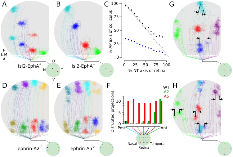
Figure 5. EphA and ephrin-A mutants.
 , Arbor development from RGCs at four retinal locations in simulated EphA knock-in experiments (Isl-EphA
 ), where 50% of RGCs had higher simulated EphA representation and thus stronger repulsion from posterior colliculus. Dual maps are formed.
  , Arbors from the same four retinal locations as in (A), but in a control (WT) retina.
  , Arborization location in colliculus as function of retinal location. The WT projection (gray) is linear, indicating a single contiguous projection of retina onto colliculus. The EphA projection is split into two contiguous maps, with blue indicating the TZ location of RGCs having upregulated EphA and black showing the locations of unaltered RGCs. Arborizations of unaltered RGCs normally targeting anterior colliculus are shifted posteriorly in simulated mutants (also visible comparing
  and B).
  , TZ location from eight points in nasal and temporal retina in simulated ephrinA2 knockouts. Arborizations are disrupted in anterior colliculus but are normal in posterior colliculus.
  , Simulated ephrinA5 knockouts. Organization is disrupted throughout the colliculus.
  , Chart showing relative number of disrupted projections in simulated WT, A2 and A5 retinas, as a function of retinal location. Axons were marked in four locations in seven dorsal-ventral bands of the retina (n = 3 simulations of each type) and the number of arbors having ectopic projections from each band was counted (maximum = 12). While ectopic projections were observed in simulated WT near collicular boundaries, the frequency and degree of these disruptions was minor in comparison to those observed in simulated ephrinA2 and A5 mutants in (D) and (E).
  ,
, Simulation results from ephrinA5 knock-out (
 ) and blocking all ephrinA/EphA mediated guidance (H). Manipulating molecular guidance on only the anterior-posterior axis can cause perturbations along the orthogonal axis, a phenomenon observed experimentally [7].

