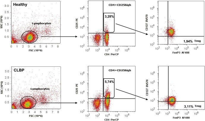
Figure 4. Gating strategy for the detection of Tregs.
PBMCs extracellular stained with PerCP labeled anti-human CD4-antibody, PE labeled anti CD25-antibody, Brilliant Violet (BV570) labeled anti CD127-antibody and intracellular stained with Alexa Fluor (AF488) labeled anti-human FoxP3-antibody. Lymphocyte population was gated from PBMCs according to forward scatter (FSC) characteristics and side scatter (SSC) characteristics (left). Gated lymphocytes were then separated in CD4+CD25high cells/T cells (middle) and CD4+CD25highCD127lowFOXP3+ cells/CD4+T cells (right, named Treg). Upper row represents the result of a healthy control with less CD4+CD25high T cells (3.28%) and less CD4+CD25highCD127lowFoxP3+ T cells (1.94%) compared to a patient with CLBP (lower row, 5.74% and 3.11%).

