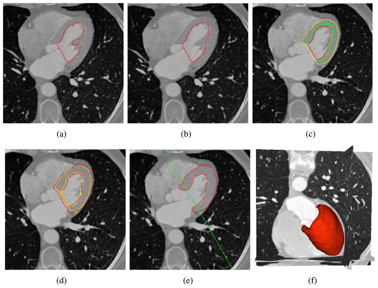
Fig. 6.
Segmentation of the myocardial wall. Initialize the endocardial surface (red) (a) before and (b) after removing papillary muscles. (c) Initialize the epicardial surface (yellow) from the initial endocarial surface (red) with a seed region (green). (d) Evolve the myocardial surfaces from the initial contours (yellow). (e) Extract the myocardial wall from endo- and epi- cardial masks by using the dividing plane (green). (f) The 3D visualization of the segmented myocardial wall.

