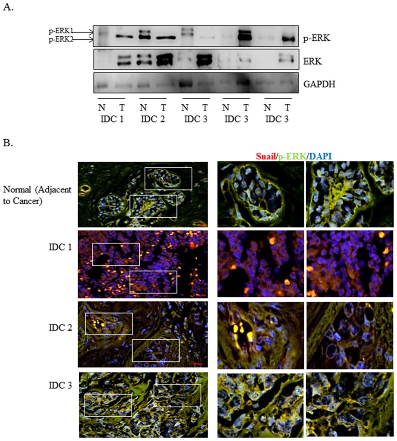
Figure 1. p-ERK expression is increased in patient tumor tissues.
(A) 10 µg of normal/tumor-matched infiltrating ductal carcinoma (IDC) grades 1–3 and lymph node metastatic patient lysates were separated using SDS-PAGE electrophoresis, then immunoblotted onto nitrocellulose. Expression of p-ERK and ERK was determined using Western blot analysis. β-actin was used as Western blot loading control. (B) Human breast cancer tissue microarray was double-labeled with p-ERK (green) and Snail (red) antibodies using immunofluorescence analysis. DAPI was used to identify the nuclei. Images were captured using Zeiss Axiovision Rel4.8 at 20× (left panel) and Apotome software at and 40× oil magnification (right panel). Results are representative of at least three independent experiments.

