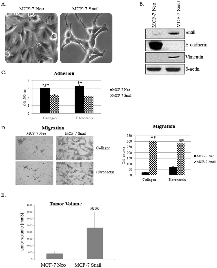
Figure 3. Snail increases EMT in vitro and tumorigenicity in vivo.
(A) Morphology images of MCF-7 Neo and MCF-7 Snail cells were captured by brightfield microscopy (100× magnification). (B) Expression of EMT markers Snail, E-cadherin, and Vimentin was analyzed using Western blot. (C) Adhesion and (D) migration assays performed on collagen I and fibronectin matrices were imaged at 10× magnification (left panel), counted and graphed (right panel). (E) MCF-7 Neo or MCF-7 Snail cells were injected subcutaneously into nude female mice (N = 6) and two weeks later, mice were sacrificed and tumor volumes measured and graphed. β-actin was utilized as a loading control for Western blot analysis. Statistical Analysis was done using paired Student's t-test, Bars, SD (**p<0.01, ***p<0.001). Results are representative of at least three independent experiments.

