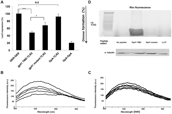
Figure 4. The gp41 TMD interacts directly with the TLR2 TMD in vitro, partially through its GxxxG dimmerization motif.
(A) GALLEX assay reveals the interaction of the gp41 TMD with the TLR2 TMD. GpA and G83I were used as positive and negative controls of interaction, respectively. (n = 3, **-p<0.005, ***-p<0.0005). (B–C) FRET analysis of the interaction between the gp41 TMD (0.1 µM) (B) and gp41 mutant (0.1 µM) (C) peptides with the TLR2 TMD peptide in LUVs. Highest line represents fluorescence of the NBD peptide in the absence of acceptor peptides. Titration with successive amounts of acceptor peptides was performed from at 1∶40, 1∶20, 1∶10 and 1∶1 acceptor∶donor ratios. (D) co-immunoprecipitation (Co-i.p.) of Rho labeled peptides together with TLR2 proteins. 1*10∧6 cells were incubated for 2 hours with the indicated peptides and then cells were lysed and the Co hours with the indicated peptides and then cells were lysed and the Co-i.p. was performed. Band detection was performed by using a fluorescence spectra-photometer scanner. Figure is a representative of 3 independent experiments.

