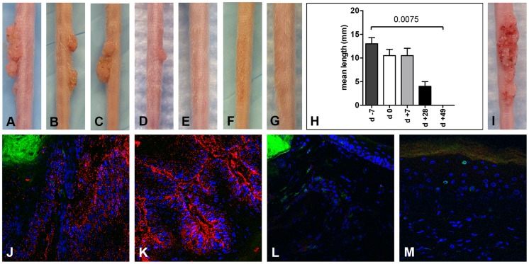
Figure 3. CD3+ T cell depletion allows papilloma formation in Cr:ORL SENCAR mice.
Infection of Cr:ORL SENCAR mice was performed on day 0 using 7.3×10¹⁰ MusPV1 virions and depletion of CD3+ T cells achieved by administration of anti-murine CD3 monoclonal antibodies for a total of 7 weeks. Efficient papilloma formation was observed when CD3+ immunodepletion was started (A) 7 days prior to, (B) on the day of, and (C) 7 days after infection. Efficiency was markedly reduced when depletion was started (D) 28 days post-infection, and no papilloma outgrowth was observed when depletion was started (E) 49 days after infection. (F) Isotype-depleted MusPV1-infected control animals and (G) MusPV1-infected control mice did not develop papillomas. (H) Comparison of tail lesions in these animals after 7 weeks of T cell depletion showed the differences in mean lesion lengths, given in mm, between the different experimental groups. Data represent the mean ± SEM of five mice/group from a representative experiment. (I) MusPV1 virions that had been extracted from lesions of CD3+ T cell depleted/MusPV1-infected Cr:ORL SENCAR mice gave rise to papillomas on the tail of an athymic NCr nude mouse after experimental transmission. (J–K) Immunofluorescent staining revealed exceptionally large amounts of MusPV1 L1 protein (red, detected with an Alexa Fluor 594-labeled secondary antibody) in the papillomatous lesions taken from MusPV1-infected Cr:ORL SENCAR mice after 7 weeks of CD3+ T cell depletion (day −7 group shown as representative). (L–M) In contrast, L1 expression was absent in skin tissues taken from isotype-depleted controls. Co-stainings of (J, L) CD4+ T cells or (K, M) CD8+ T cells in the tissues were performed using Alexa Fluor 488-labeled anti-CD4 or anti-CD8 antibodies (green), respectively, and confirmed the absence of T cells in the depleted animals. Infection of all animals was performed on day 0 using 7.3×10¹⁰ MusPV1 virions.

