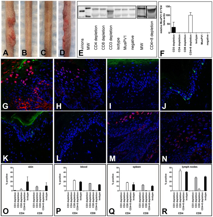
Figure 5. CD3+, but not single CD4+ or single CD8+ T cell depletion allows papilloma formation in C57BL/6 mice.
Wild-type C57BL/6 mice were infected with 5.3×10¹⁰ MusPV1 virions, and the depleted state maintained for a total of 7 weeks. After this period papillomas developed in (A) CD3-depleted (n = 14), but not in (B) single CD4-depleted (n = 5) and (C) single CD8-depleted (n = 5) C57BL/6 mice. (D) Combined CD4+8 depletion allowed papilloma outgrowth in C57BL/6 mice (n = 10). (E) MusPV1 L1 protein at the expected size of 55–60 kD was found by Western Blot in crude skin tissue lysates prepared from CD3 and combined CD4+8-depleted C57BL/6 mice, but was absent in tissue lysates from single CD4-depleted or single CD8-depleted animals. L1 protein was also undetectable in MusPV1-infected, non-depleted and mock-infected littermates. One representative per group is shown. The molecular weight marker (MW) is depicted on the left side and 60 kD and 50 kD markers are visible; purified MusPV1 virions served as controls. (F) MusPV1-specific E1∧E4 spliced transcripts relative to endogenous beta-actin were detected at high levels in skin tissues harvested at 6 weeks post-infection (corresponding to 7 weeks of depletion) from CD3 and combined CD4+8-depleted C57BL/6 mice. E1∧E4 spliced transcripts were undetectable in tissues from CD4-depleted, CD8-depleted animals, and appropriate controls. Immunofluorescent staining showed the presence of MusPV1 L1 protein (red, visualized using an Alexa Fluor 594-labeled secondary antibody) in skin tissues harvested after the 7 week depletion period from (G,H) CD3-depleted and (M,N) combined CD4+8-depleted C57BL/6 mice. Consistent with the lack of E1∧E4 spliced transcripts or L1 protein expression in crude lysates, L1 protein was undetectable in tissues from (I,J) single CD4-depleted and (K,L) single CD8-depleted. C57BL/6 mice. Co-staining of (G, I, K, M) CD4+ T cells with a directly Alexa Fluor 488-labeled anti-CD4 antibody (green) or co-staining of (H, J, L, N) CD8+ T cells confirmed the efficient and specific immunodepletion in these tissues. Flow cytometry analyses of CD4+ (left side) and CD8+ (right side) T cells in (O) skin tissues, (P) blood, (Q) spleen and (R) draining lymph nodes of T cell depleted C57BL/6NCr mice further corroborated maintenance of the depleted state in each compartment.

