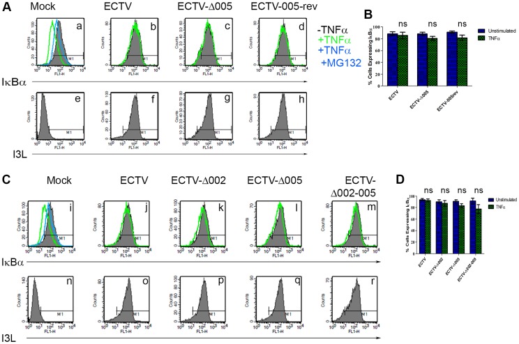
Figure 7. ECTV-Δ005, ECTV-Δ002 and ECTV-Δ002-005 inhibit TNFα induced IκBα degradation.
(A) HeLa cells were mock-infected or infected with ECTV, ECTV-Δ005 or ECTV-005-rev at a MOI of 5. (C) Alternatively, HeLa cells were mock-infected or infected with ECTV, ECTV-Δ002, ECTV-Δ005 or ECTV-Δ002-005 at a MOI of 5. (A and C) At 12 hours post hours post-infection, cells were stimulated with 10 µM MG132 for 1 hour and hour and/or 10 ng ng/ml TNFα for 20 minutes. Cells were harvested, fixed and permeablized, followed by staining with anti minutes. Cells were harvested, fixed and permeablized, followed by staining with anti-IκBα and anti-I3L. Samples were subjected to flow cytometry, IκBα (A panels a–d, and C panels i–m) or I3L (A panels e–h and C panels n–r) are measured along the x-axis. (B and D) The percentage of cells expressing IκBα were measured and plotted as the average of three independent experiments with SEM.

