
Figure 1. Morphological Evidence of PG Inhibiting Dox Induced Apoptosis.
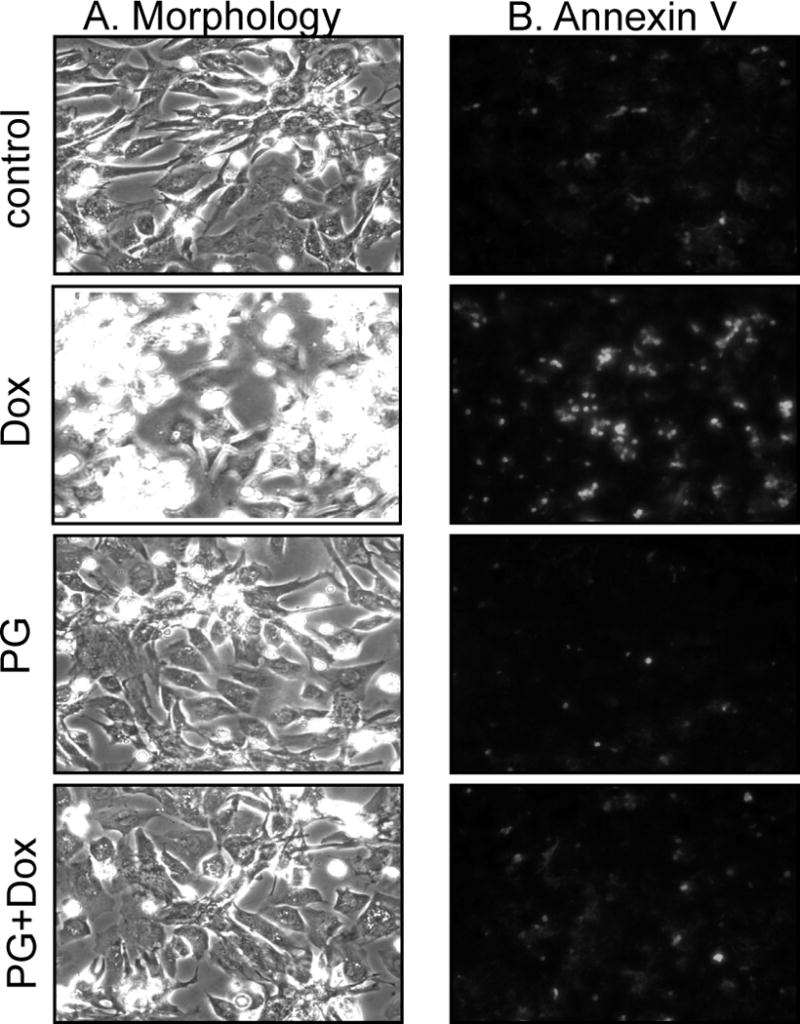
Primary cultured cardiomyocytes were pretreated with 10 μM PG for 24 hrs and then treated with 0.8 μM Dox in the absence or presence of 10 μM PG. At 24 hrs after Dox treatment, the morphology was recorded under a phase contrast microscope. Detached cells were combined with adherent cells for Annexin V-Fluo staining for recording under a fluorescent microscope.