
Figure 3. Expression of PR in Cardiac Tissue or Cardiomyocytes.
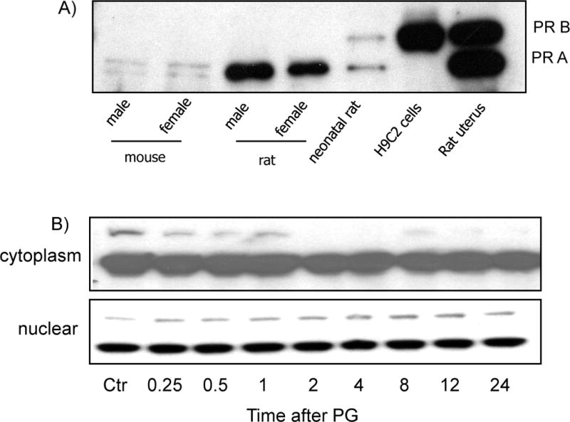
Cardiac tissue was collected from 8 weeks old male or female C57BL6 mice or Sprague Dawley rats by quick frozen in liquid nitrogen. The uterus tissue from a pregnant rat was collected immediately after birth to serve as a positive control. Animal tissues were grinded in liquid nitrogen bath for dissolving in lysis buffer for Western blot analyses (A). Primary cultured cardiomyocytes were treated with 10 μM PG and harvested at indicated time for isolation of cytoplasmic or nuclear fractions (B). An equal amount of proteins (20 μg/lane) was loaded for Western blot analyses.