
Figure 7. PG Causes An Increase in the Rate of Bcl-xL Transcription.
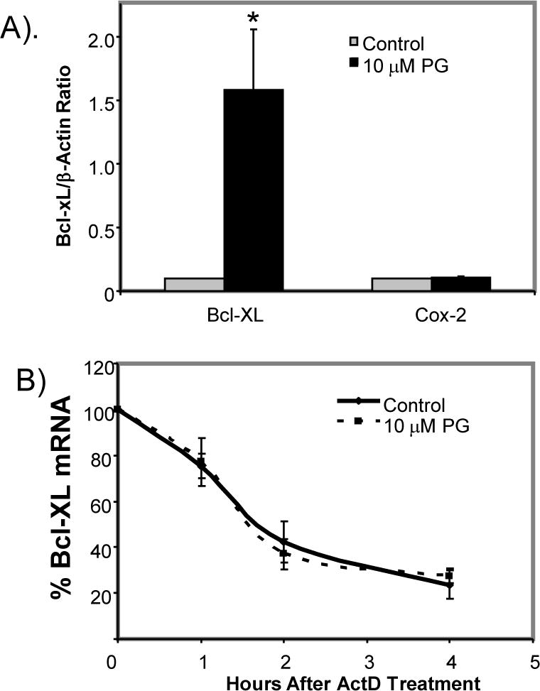
Primary cultured cardiomyocytes were treated with 10 μM PG for 24 hrs before being harvested for nuclear run-on assay (A) or measurements of Bcl-xL mRNA stability by addition of 5 μg/ml actinomycin D (ActD) over indicated time (B). The relative Ct method was used to calculate mRNA levels with normalization to β-Actin (A). The level of Bcl-xL was set to 100 at 0 time point for determining Bcl-xL mRNA stability (B). The data represent average ± standard deviations of three independent experiments. An asterisk (*) indicates p<0.05 when the treated group was compared to control using Student’s t test.