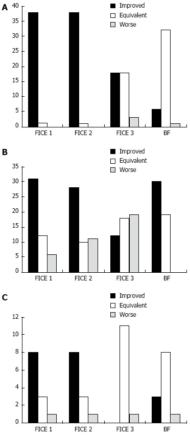
Figure 2.

Delineation. A: Of angioectasias with all different settings of virtual chromoendoscopy: comparison with conventional white light; B: Of ulcers or erosions with all different settings of virtual chromoendoscopy: comparison with conventional white light; C: Of villous edema or atrophy with all different settings of virtual chromoendoscopy: comparison with conventional white ligh.
