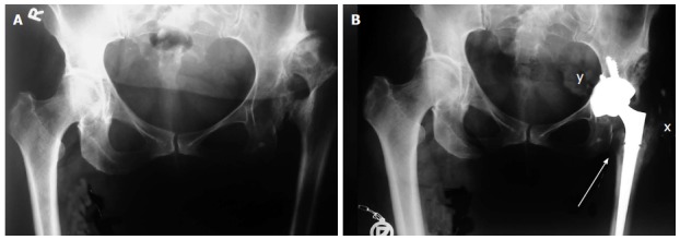
Figure 6.

X-rays of patient with Crowe type 4 dysplasia on the left side and normal hip on right side. A: Preoperative X-ray with secondary osteoarthritis due to dysplasia, neoacetabulum formed superolaterally from original, true acetabulum and significant leg length discrepancy; B: Postoperative X-ray with implanted uncemented acetabular cup and femoral stem. Acetabular cup is protruding beyond the Kohler’s line inside the pelvis (marked with y) and secured with 3 additional screws. Lesser trochanter is brought distally to the normal level so there is no leg length discrepancy postoperatively (marked with a single arrow). Modified direct lateral approach was used and posterior part of the gluteus medius and vastus lateralis together with the external rotators were detached with the chisel on a thin flake of bone, now they are completely attached and healed to greater trochanter (marked with x).
