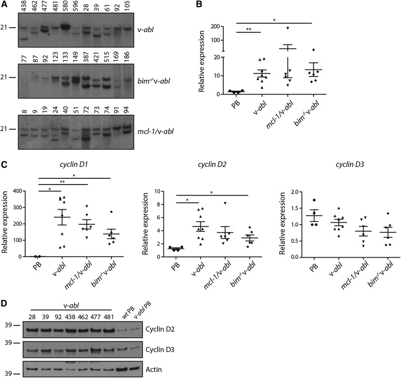
Figure 6.
Activation of myc and cyclin D genes in v-abl plasmacytomas. (A) Southern blot analysis of EcoRI-digested DNA (20 μg) from v-abl, bim−/−v-abl and mcl-1/v-abl tumors using a c-myc exon 3 probe. (B) Real-time PCR analysis of cDNA prepared from frozen tumor samples and PBs generated in vitro. All v-abl plasmacytomas express high levels of c-myc mRNA, including those for which c-myc translocations were not apparent in A. Expression was normalized to HMBS, and the data are expressed relative to PBs. Mean ± SEM, n = 6 to 8 independent tumors. *P < .05, **P < .01, Student t test. (C) Real-time PCR analysis of cyclin D1, D2, and D3 mRNA in v-abl plasma cell tumors and PBs (generated in vitro). Expression was normalized to HMBS, and the data were expressed relative to v-abl PBs. Mean ± SEM, n = 6 to 8 independent tumors. *P < .05, **P < .01, Student t test. (D) Western blot analysis of cyclin D2 and D3 protein expression in v-abl tumors (whole tissue) and PBs (generated in vitro).

