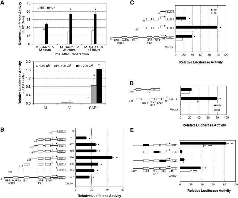
Figure 1.
The 5′-region of the SAR1 gene exhibits promoter activity that is inducible by HU and contains intact Elk-1/NF-κB–binding sites that are required for basal and HU-induced SAR1 promoter activity. (A) Top panel, −977 to +49 genomic region containing the predicted promoter region was cloned into the pGL3 basic luciferase vector and transfected into K562 cells with (+) or without (−) 2 days of 100 μM HU pretreatment. The level of promoter activity in K562 cells was evaluated 12, 24, and 48 hours after transfection. Bottom panel, SAR1 reporter construct was transfected into CD34+ cells with 2 days of 0, 100, or 200μM HU pretreatment at day 6 of differentiation. The level of promoter activity in CD34+ cells was evaluated 24 hours after transfection. Mock-, SAR1 reporter construct–, and vector control–transfected cells are indicated as M, SAR1, and V, respectively. *P < .05 vs HU untreated cells. (B) Deletions of the SAR1 promoter region were constructed and transfected into K562 cells, then promoter activity measured 24 hours after transfection. Left side, Schematic diagram of deletions with transcription factor–binding sites indicated. *P < .001 vs vector control–transfected cells. (C-D) Reporter constructs containing different fragments of the SAR1 promoter were transfected into K562 cells with (+) or without (−) 2 days of 100 μM HU pretreatment. Left side, Schematic diagram of deletion constructs. Luciferase activities were measured 24 hours after transfection. *P < .05 vs HU untreated cells. (E) Mutation analysis of the effects of Elk-1, EKLF, and Elk-1/NF-κB transcription factor binding sites on SAR1 promoter activity. Left side, Schematic diagram of wild-type and mutant reporter constructs. Luciferase activities were measured 24 hours after transfection into K562 cells with (+) or without (−) 2 days of 100 μM HU pretreatment. □, Wild-type transcription factor–binding sites; ▪, mutated transcription factor–binding sites. **P < .001 vs vector control–transfected cells without HU treatment; *P < .05 vs HU untreated cells. The level of promoter activity was evaluated by measurement of the firefly luciferase activity relative to the internal control Renilla luciferase activity using the Dual Luciferase Assay system. Error bars represent SD of the mean of 3 independent experiments. SD, standard deviation.

