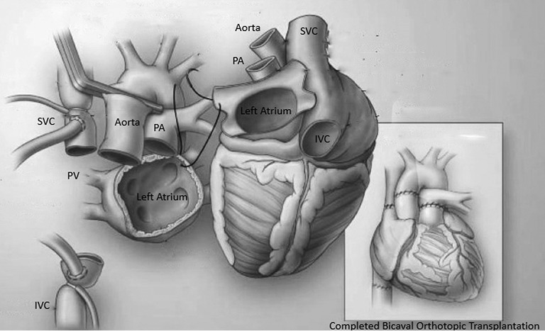
Figure 5.

An illustration of the recipient mediastinum and the donor heart with the first stitch at the level of the donor left atrial appendage and recipient left superior pulmonary vein. The illustration on the right showed the completion of a bicaval orthotopic heart transplantation. PA, pulmonary artery; PV, pulmonary vein; SVC, superior vena cava; IVC, inferior vena caca.
