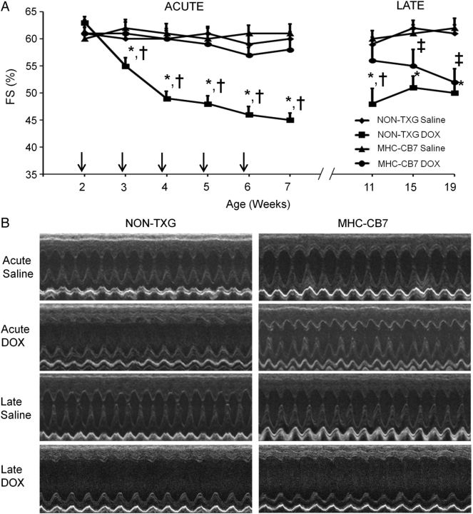
Figure 1.
Cardiac function in saline- and DOX-treated juvenile mice in the acute and late stages. (A) FS in saline- and DOX-treated NON-TXG and MHC-CB7 mice in the acute and late stages. X-axis indicates the age of the mice at analysis; vertical arrows indicate the age at DOX injection. *P < 0.05 for DOX-treated NON-TXG vs. saline-treated NON-TXG mice, †P < 0.05 for DOX-treated NON-TXG vs. DOX-treated MHC-CB7 mice, ‡P < 0.05 for DOX-treated MHC-CB7 vs. saline-treated MHC-CB7 mice. (B) Representative short-axis echocardiograms from saline- and DOX-treated NON-TXG and MHC-CB7 mice in the acute and late stages.

