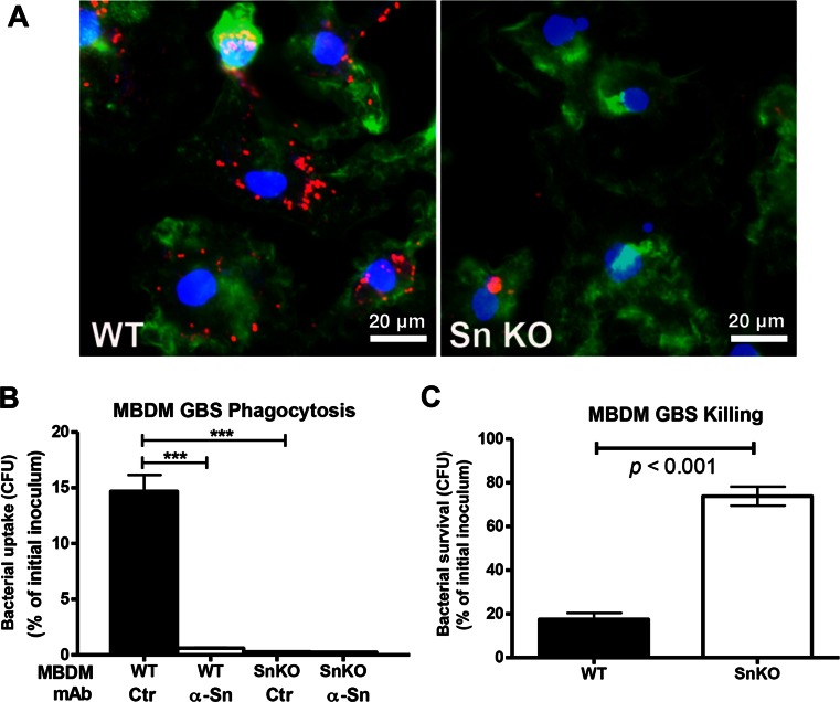
Fig. 2.
Sn contributes to phagocytic and bactericidal activity against GBS. a Confocal microscopy images show significantly reduced phagocytosis of GBS in the Sn-deficient (Sn KO) MBDMs. Macrophages were infected with GBS at MOI of 5 for 30 min and washed extensively before staining cells with actin. Red indicates GBS. Green indicates actin. Blue indicates nucleus. b Quantitative phagocytosis analysis. MBDMs were infected with GBS in the presence or absence of Sn-neutralizing mAb, followed by addition of antibiotics to only recover and enumerate intracellular bacteria CFU. c Survival of GBS upon co-incubation with MBDMs. Macrophages were infected with GBS at MOI of 0.2, and surviving GBS was enumerated 1 h post-infection by serial plating. Difference between two groups was calculated one-way ANOVA with Tukey’s multiple comparison test (b) or by unpaired t test (c). ***P < 0.001. Representative image (a) and data (b and c) were shown from three independent experiments

