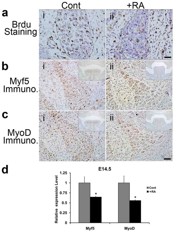
Figure 2. BrdU cell proliferation assay and immunohistochemistry (IHC) assay of the genioglossus at E14.5, and qRT-PCR analyses of Myf5 and MyoD expression at E14.5.
(a): Cell proliferation assay. At E14.5, the numbers of BrdU positive cells/cm2 in the genioglossus of +RA mouse fetuses (ii), decreased to 35.3% of those observed in controls (i) (n = 3 mice per group, P < 0.01). The boundary of genioglossus was delineated by the dotted line. (b)–(c): IHC assay. (b) Myf5 protein level was apparently lower in the genioglossus of the +RA mouse fetus (ii) than control (i). Bar = 40 μm. (c) MyoD protein level in the genioglossus of the +RA fetus (ii) was lower than control (i). Bar = 40 μm. Upper right inserts in (b ii and c iii) show images at lower magnifications, Bar = 100 μm. (d) qRT-PCR analyses. The mRNA levels of Myf5 and MyoD decreased significantly in the +RA fetus as compared to the controls (n = 5, P < 0.05). Cont: control mouse fetus; +RA: RA-exposed mouse fetus.

