
Figure 3. Phosphorylation assay.
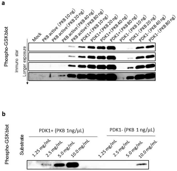
(a) GSK3peptide fusion proteins were separated by 15% SDS–PAGE and subjected to a Western blotting analysis using a GSK3β/α antibody. Commercially available PKBα is indicated by “PKB active”, PKB with PDK1 co-expression as “PDK1+”, and PKBα without PDK1 co-expression as “PDK-”. Four different panels that show the bands visualized by different exposure time are shown. (b) Western blots showing dose related variations in phosphorylation by GSK3peptide fusion proteins. (a)(b) The regions of interest are cropped.