
Fig. 2. Studying osteoclastogenesis using high-content imaging.
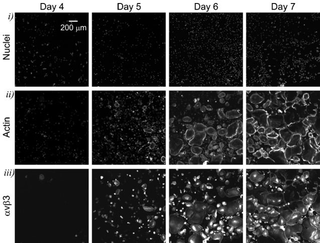
Human osteoclast precursors were cultured with MCSF (33 ng/ml) and RANKL (33 ng/ml) for 4–7 days, fixed and stained with Hoechst for nuclei, phalloidin for actin as a cytoplasmic marker, and for αvβ3 integrin as an osteoclast marker. Representative images demonstrate temporal changes in the appearance of nuclei (top) and actin (middle), and emergence of αvβ3 integrin (bottom) during osteoclast differentiation. Scale bar: 200 µm (applies to all images).