
Figure 1. Benchmarking myogenic differentiation in human eRMS.
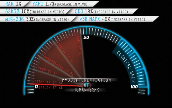
Representative interventions reported as percentage increase of MHC positive cells*in vitro or in vivo(Barlow et al., 2006; Chen et al., 2014; Puri et al., 2000; Taulli et al., 2009; Wegorzewska et al., 2003). Corresponding targets are noted. For consistency, only studies of the prototypic RD cell line (generated in 1968) are included. Some of these pathways may be interlinked (e.g., GSK3β and YAP1 have been reported to be co-associated on the Axin scaffold, regulating β-catenin and YAP1 signaling in parallel).*In the case of RAR, MHC was not done but the authors reported no differentiation by morphology or by Troponin-T immunocytochemistry in response to retinoic acid. Illustration by Nick Escobar.