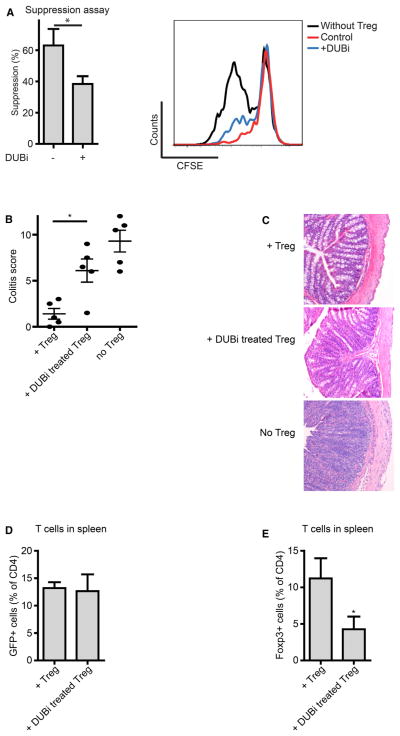
Figure 1. Ubiquitination Modulates Treg Cell Function.
(A) Sorted human CD4+CD25highCD127low Treg cells were pretreated with 10 μM DUBi for 1 hr, washed, and cocultured with CFSE-labeled PBMCs in anti-CD3-coated wells for 4 days. CFSE dilution of CD4+ cells was analyzed by flow cytometry.
(B) CD4+CD45RBhigh cells were injected into immunodeficient mice. Sorted GFP+ Treg cells from Foxp3-GFP promoter mice were pre-incubated with 10 μM DUBi for 1 hr and injected 3 weeks later. Mice were sacrificed 3 weeks after Treg cell administration. Sections of the colon were analyzed and scored (five mice per group).
(C) Representative hematoxylin- and eosin-stained tissue slides of the colon.
(D) Analysis of CD4+GFP+ cell numbers in the spleen.
(E) Percentage of Foxp3+ CD4+ T cells in the spleen. Data shown are representative of at least three independent experiments, *p<0.05.
Data are represented as mean + SEM. See also Figure S1.

