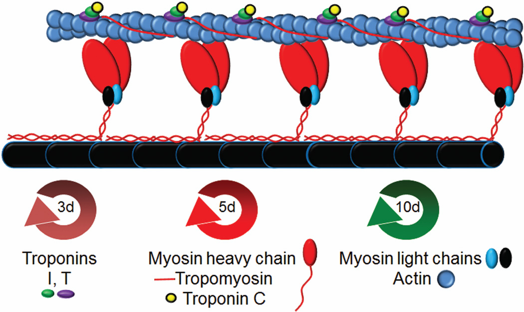
Figure 1.
Schematic representation of the structure of the cardiac sarcomere. The sarcomere is made up of the thin filament containing actin, troponin, and tropomyosin and the thick filament containing myosin, myosin light chains, and titin (black) along with myosin binding protein C (not depicted). Troponin interacts with tropomyosin and actin in 1:1:7 stochiometry. Myosin motor domains (red) interact with actin in a calcium dependent manner to produce force. Turnover half-lives of the depicted sarcomeric proteins are presented below (d=days).

