Figure 4.

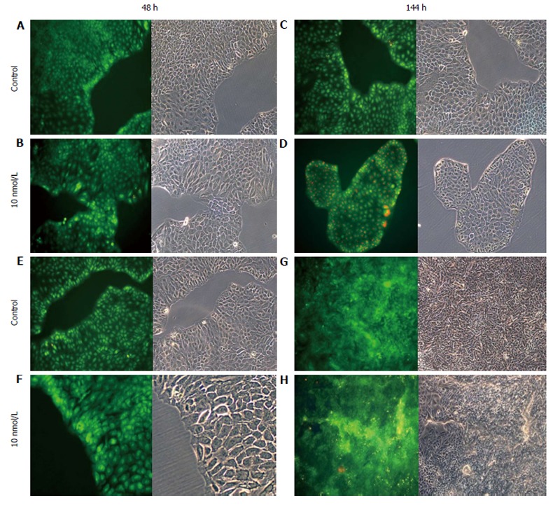
Treatment of cells with 17β-E2. A-D: HCT8-β8-expressing cells; or E-H: HCT8-pSV2neo-expressing cells were treated with 10 nmol/L 17β-E2 for 48 h (A, B, E, F) or 144 h (C, D, G, H) and stained with acridine orange. Nuclei and mitochondria appear green, whereas lysosomes appear red-orange under fluorescence, adjacent to corresponding phase contrast images (magnification × 20).
