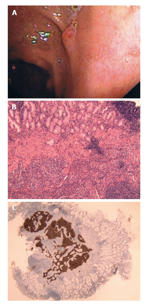
Figure 1.

A 65-year-old female with a history of possible inflammatory bowel disease presented for evaluation of epigastric pain and occasional hematochezia. A: Patient 1, neuroendocrine (carcinoid) tumors as duodenal nodule at endoscopy; B: Solid growth pattern with organoid architecture and bland monotonous cells with lack of significant atypia and increased mitoses. H and E, × 10; C: Neoplastic neuroendocrine cells show diffuse positivity for Chromogranin. Chromogranin, × 20.
