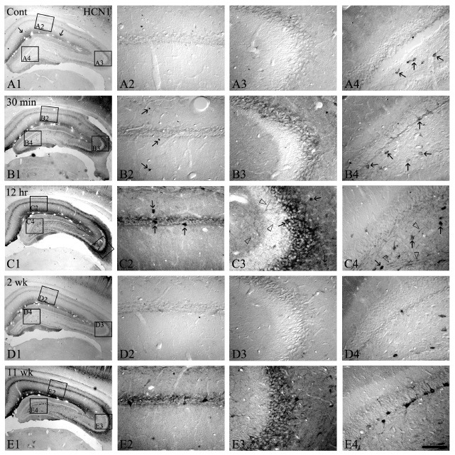
Fig. 1. The HCN1 expressions in the hippocampus following pilocarpine-induced SE. HCN1 immunoreactivity is selectively detected in the stratum lacunosum-molecule and some hilar neurons (arrows in panel A1 and A4). However, HCN1 immunoreactivities at 30 min-12 hrs following SE are significantly enhanced in CA1-3 and the stratum lacunosum-molecule (B1-B3 and C1-C3). In addition, HCN1-positive interneurons and hilar neurons are increased (arrows and open arrows in panel B2, B4, C2, and C4). Nevertheless, at 2 weeks following SE, HCN1 immunoreactivities are down-regulated to levels similar to the control (D1-D4). At 11 weeks after SE, HCN1 expression in all hippocampal regions and its immunoreactive interneurons is re-enhanced, similar to 12 hrs following SE (E1-E4). Bar = 280 μm (panels A1, B1, C1, D1, and E1), 50 μm (panels A2-A4, B2-B4, C2-C4, D2-D4, and E2-E4).