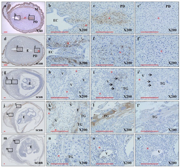
Fig. 1. Differential expression of KAI1 at the feto-maternal interface during different embryonic periods. Paraffin sections of whole uteri and embryos of C57BL/6 mice at E5.5 (a), E7.5(d), E9.5(g), E11.5(j), and E13.5(m) were prepared to detect KAI1 protein with rabbit polyclonal anti-KAI1 antibody as described in “Materials and Methods.” Sections were treated with anti-KAI1 antibody (a, d, g, j, and m). Images b, c, e, f, h, i, k, l, n, and o are higher-magnification images corresponding to the small rectangles in a, d, g, j, and m, respectively. The sections of c’, f’, i’, l’, and o’ were stained with normal IgG as an internal control of c, f, i, l, and o, respectively. E, embryo; EC, endometrial cavity; M, myometrium; PD, predecidua; T, trophoblast; TG, trophoblast giant cell; V, Uterine spiral artery; *, decidua; →, glycogen trophoblast cell. Scale bar indicates 200 μm.

