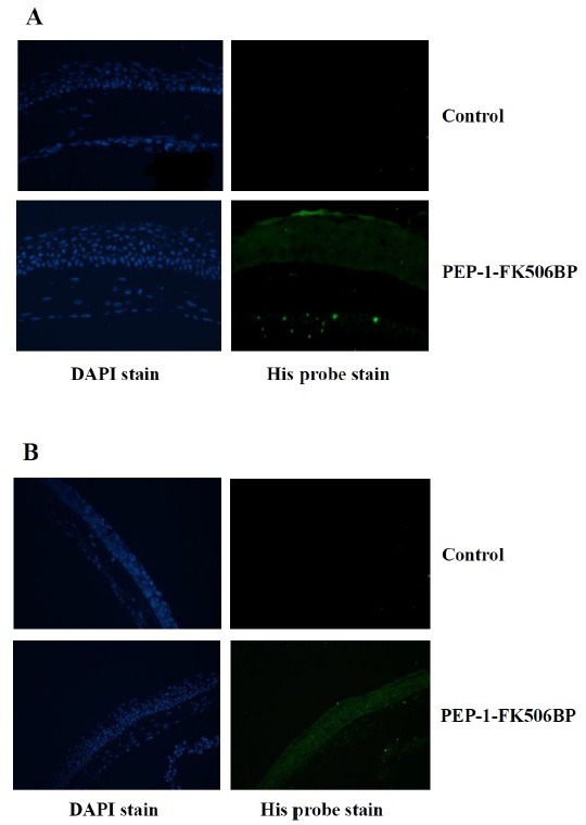
Fig. 2. Transduction of PEP-1-FK506BP in cornea and conjunctiva. Representative images of the immunofluorescent staining of PEP-1-FK506BP-treated mice cornea (A) and conjunctiva (B) using anti 6-His antibody. The staining was strong in PEP-1-FK506BP-treated mice compared to that in control mice.

