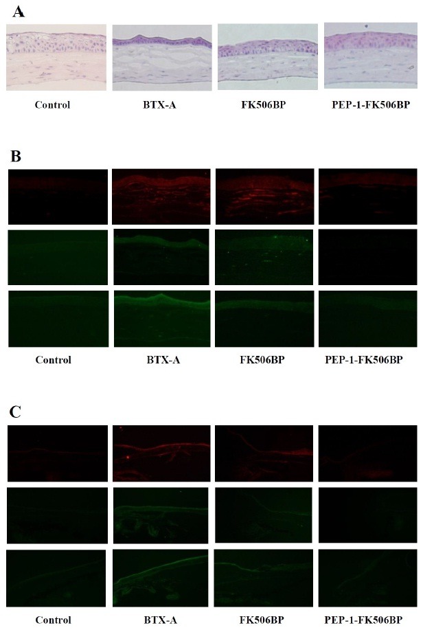
Fig. 4. Anti-inflammatory effects of PEP-1-FK506BP in corneal and conjunctival epithelia. (A) Images of H&E staining after BTX-A-injection, FK506BP-treatment, and PEP-1-FK506BP-treatment. Immunofluorescent staining of corneal (B) and conjunctival epithelia (C) using indicated antibody. The staining for cytokines was strong in BTX-A-injected mice. The PEP-1-FK506BP treated mice group demonstrated significantly decreased cytokine expression compared with BTX-A or FK506BP treated groups.

