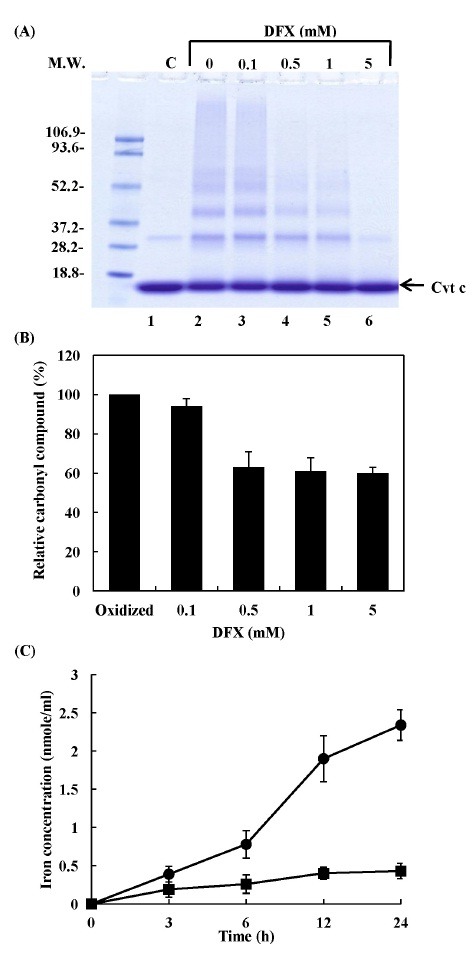
Fig. 3. Effects of iron chelator on salsolionl-mediated cytochrome c aggregation and carbonyl compound formation and salsolinol-mediated iron release from cytochrome c. 20 μM cytochrome c was incubated with 0.5 mM salsolinol in 10 mM phosphate buffer (pH 7.4) at 37℃ for 24 h in the presence of DFX. (A) The pattern of protein bands was analyzed via SDS-PAGE. Lane 1, cytochrome c control; lane 2, oxidized cytochrome c (without DFX); lane 3, 0.1 mM DFX; lane 4, 0.5 mM DFX; lane 5, 1 mM DFX; lane 6, 5 mM DFX. (B) The formation of carbonyl compounds was determined by spectrophotometry. (C) The reaction mixture contained 20 μM cytochrome c and 0.5 mM salsolinol in 10 mM phosphate buffer at pH 7.4, and was incubated for various periods of time as: cytochrome c alone (■); cytochrome c plus salsolinol (●). Free iron ions were determined with a colorimetric reagent using bathophenanthroline sulfonate.