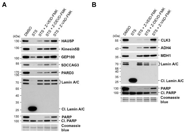
Fig. 3. Cleavage of caspase-6 substrate candidates was confirmed by in vivo cleavage assay. HeLa cells were treated with 1 μM STS for 24 hr in the presence or absence of the 100 μM caspase inhibitors (Z-VEID-FMK; caspase-6 inhibitor, Z-DEVD-FMK; caspase-3/-7 inhibitor, Z-VAD-FMK; pan-caspase inhibitor) pretreated to cells for 3 hr. After 24 hr, cells were harvested and lysed. Cell lysates were analyzed by immunoblotting with the specific antibodies against (A) HAUSP, Kinesin5B, GEP100, SDCCAG3, PARD3, Lamin A/C (Cl, cleaved Lamin A/C), and PARP (Cl, cleaved PARP); and (B) CLK3, ADH4, MDH1, Lamin A/C, and PARP. A coomassie blue stain of PVDF membrane was the loading control. The representative blot of at least two independent experiments is shown.

