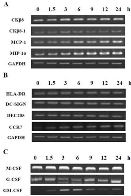
Fig. 1. mRNA expression of GM-CSF was affected by MTB. THP-1 cells were treated with PMA (100 nM) for 48 h and were incubated in the presence of MTB for the indicated times (0, 1.5, 3, 6, 9, 12, 24 h). cDNA were prepared from total RNA of infected cells, and was subjected to PCR to amplify (A) chemokines (CKβ8, CKβ8-1, MCP-1, MIP-1α), (B) DC markers (HLA-DR, DC-SIGN, DEC205, CCR7), and (C) colony stimulating factors (M-CSF, G-CSF, GM-CSF). The PCR products were resolved by 1.8% agarose gel. GAPDH was used as an internal control.

