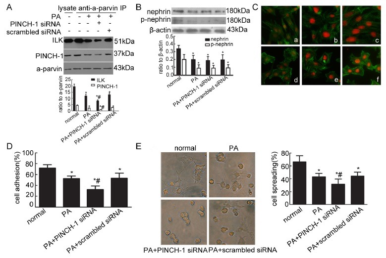
Fig. 2. Knockdown of the PIP complex resulted in the reorganization of the cytoskeleton, and down-regulated podocyte adhesion and spreading. (A) ILK and PINCH-1 were immunoprecipitated by an α-parvin antibody in cultured normal podocytes and podocytes stimulated by PA with/without PINCH-1 siRNA or scrambled siRNA transfection. The immunoprecipitates were analyzed by Western blot with antibodies of ILK, PINCH-1 and α-parvin. *P < 0.05 compared with the normal group; #P < 0.05 compared with the groups stimulated by PA with/without scrambled siRNA. (B) Nephrin and phosphorylated nephrin were detected by Western blot in different groups. *P < 0.05 compared with the normal group; #P < 0.05 compared with the groups stimulated by PA with/without scrambled siRNA. (C) Stress fiber of the cytoskeleton was detected by immunofluorescence with FITC-phalloidin. Original magnification ×400. (a) Normal podocytes; (b) podocytes transfected with PINCH-1 siRNA; (c) podocytes transfected with scrambled siRNA; (d) podocytes stimulated by PA; (e) podocytes stimulated by PA with PINCH-1 siRNA transfection; (f) podocytes stimulated by PA with scrambled siRNA transfection. (D) Cell adhesion was measured with spectrophotometry. *P < 0.05 compared with the normal group; #P < 0.05 compared with the PA or scrambled siRNA group. (E) Cell spreading was detected and counted under inverted microscope. Original magnification ×400. *P < 0.05 compared with the normal group; #P < 0.05 compared with the PA or scrambled siRNA group.

