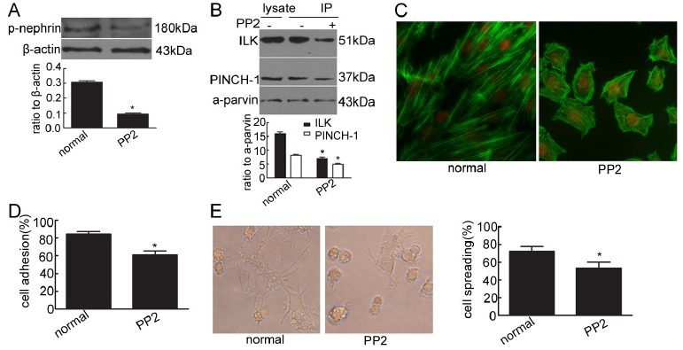
Fig. 4. Effect of phosphorylated nephrin on podocyte cytoskeleton, adhesion and spreading in the physiological state. (A) Phosphorylated nephrin was detected by Western blot with antibody against phosphorylated nephrin (pY1217) in cultured normal podocytes and podocytes stimulated by the Src family kinase inhibitor (PP2). *P < 0.05 compared with the normal group. (B) ILK and PINCH-1 were immunoprecipitated by an α-parvin antibody in cultured normal podocytes and podocytes stimulated by PP2. The immunoprecipitates were analyzed by Western blot with antibodies against ILK, PINCH-1 and α-parvin. *P < 0.05 compared with the normal group. (C) Stress fiber of the cytoskeleton was detected by immunofluorescence with FITC-phalloidin. Original magnification ×400. (D) Cell adhesion was measured with a spectrophotometer. *P < 0.05 compared with the normal group. (E) Cell spreading was detected and counted under an inverted microscope. Original magnification ×400. *P < 0.05 compared with the normal group.

