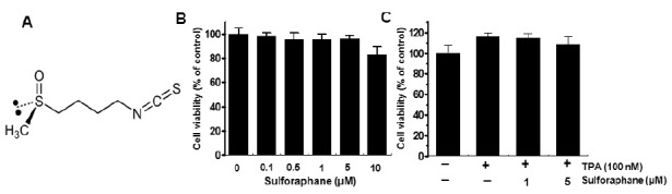
Fig. 1. Structure of sulforaphne and effect of sulforaphane on cell viability of MCF-7 viability. Chemical structure of sulforaphane (A). To examine the cytotoxicity of sulforaphane, cells were cultured in 96-well plates up to a confluency of 70%, and various concentrations of sulforaphane were added to cells for 24 h (B). MTT assay was used to detect the viability of the cells. Cells were pretreated with 1 μM and 5 μM sulforaphanes for 1 h, before incubation with 100 nM TPA for 24 h (C). The optical density of the control was regarded as 100%. Data points are the mean ± SEM of three independent experiments.