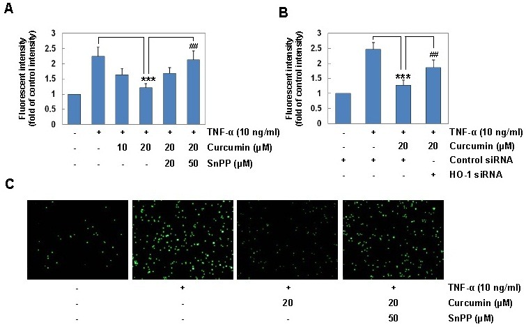
Fig. 3. HO-1 induction mediates the inhibitory effect of curcumin on TNF-α-induced monocyte adhesion in HaCaT cells. (A) HaCaT cells were incubated with 20 μM curcumin for 6 h in the absence or presence of SnPP, and then exposed to TNF-α (10 ng/ml) for 12 h. HaCaT cells were co-cultured with calcein-AM-labeled THP-1 monocytes for 1 h. The calcein-AM fluorescent intensity was measured by an ELISA plate reader. (B) HaCaT cells transfected with control or HO-1 siRNA were incubated with 20 μM curcumin for 6 h, and stimulated with TNF-α for 12 h. HaCaT cells were co-cultured with calcein-AM-labeled THP-1 monocytes for 1 h. The calcein- AM fluorescent intensity was measured by an ELISA plate reader. Results are means ± SD. Statistical significance: ***P <0.001 compared to the TNF-α alone, ##P < 0.01 compared to the TNF-α and curcumin. (C) Microphotographs were obtained using fluorescence microscopy (original magnification, ×40).

