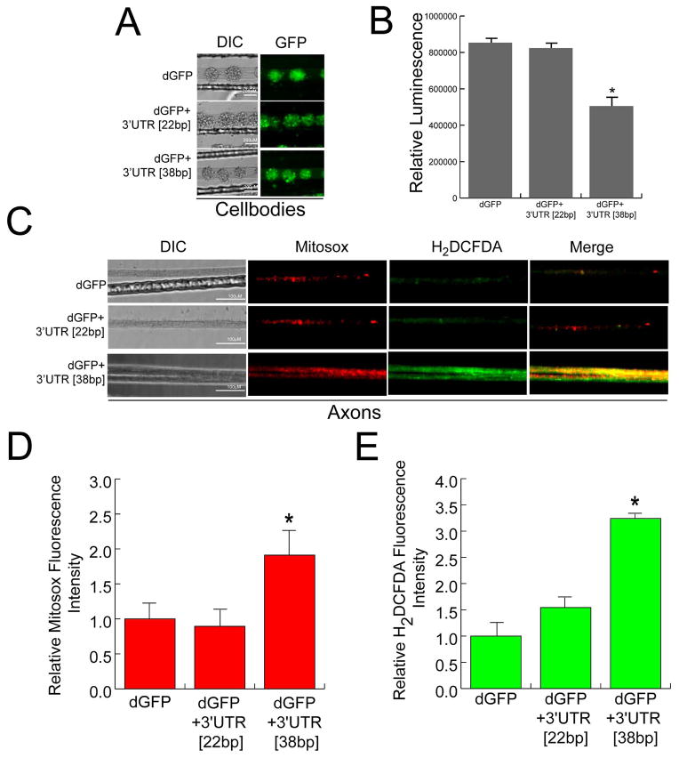
Figure 2.
Over-expression of dGFP mRNA containing the 38bp COXIV zipcode decreases local ATP levels in axons and alters axonal ROS production. SCG neuron cell-bodies (6DIV) were transfected with dGFP+3′UTR [38bp], dGFP+3′UTR[22bp], or dGFP constructs (3ug each). (A) Representative DIC and fluorescence images showing the expression of dGFP contructs in the transfected cell-bodies. (B) Total axonal ATP levels were measured in distal axons 48 h after transfection of SCG neurons using the CellTiter-Glo Luminescent Cell Viability Assay from Promega (see Methods). Values are plotted as relative luminescence units. (C) Intra-axonal reactive oxygen species (ROS) levels were measured in distal axons 24 h after transfection of cell-bodies by fluorescence microscopy using Carboxy-H2DCFDA (green) and Mitosox (red). Fluorescence intensity was quantified using ImageJ, and fluorescence levels for Mitosox (D) and Carboxy-H2DCFDA (E) are indicated as relative fluorescence units (RFU). Data are the mean ± SEM from the measurement of 35–45 axons. One-way ANOVA, *, p < 0.01.

