Figure 1.
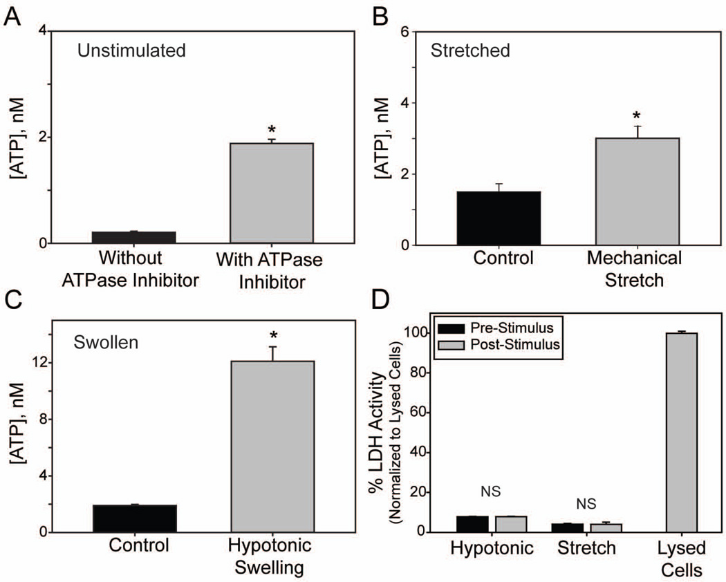
Mechanical stretch and hypotonic swelling of astrocytes releases ATP. (A): Comparison of basal extracellular ATP concentrations from non-stimulated (control) cells in the absence or presence of the ecto-ATPase inhibitors β,γ-methylene ATP and ARL67156 (100µM each; n=385, 147 respectively) (B): Effects of mechanical stretch (5% equibiaxial strain, 0.3 Hz for 2 min) on extracellular ATP concentrations. ATP levels were determined from 100µl samples taken before and after cells were stretched, n=4 each. (C): Effects of hypotonic swelling (50% dH2O) on extracellular ATP concentrations measured between 1.5–2.5min after addition of hypotonicity, n=140. All solutions in (B) and (C) had 100µM β,γ-methylene ATP and ARL67156. (D): Measurement of extracellular lactose dehydrogenase (LDH) as an index of cell lysis. LDH activity was measured before and after stimuli (hypotonic swelling or mechanical stretch). NS = no significant. Levels expressed as % of that measured from lysed cells; n=20 for hypotonic, 4 for stretch and 21 for lysed cells. *p<0.05, unpaired Students’ t-test in A,C; paired in B,D.

