
Figure 4.
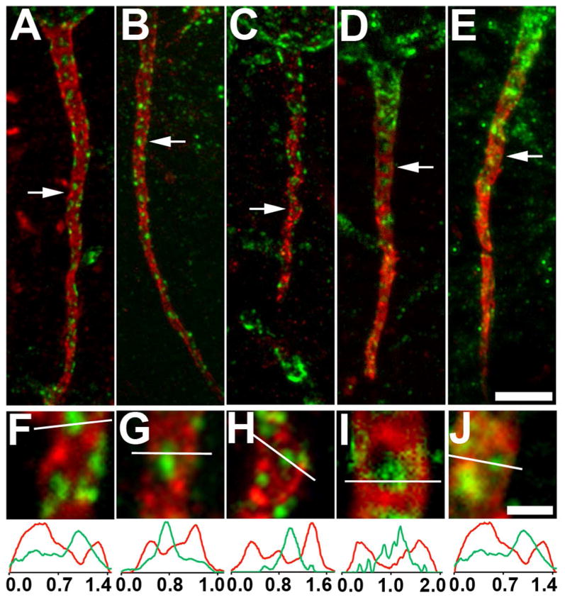
Kv2.1 is localized at AnkG-deficient sites on the AIS of layer 5 neocortical pyramidal neurons in different mammalian species. Sections double immunofluorescence labeled for Kv2.1 (green) and AnkG (red). Images were obtained from neocortical neurons in the brains of different mammalian species: A,F: rat; B,G: ferret; C,H: monkey; D,E,I,J: human. Arrows in panels A-E correspond to the location of the midpoint of the 4X enlarged insets shown as panels F-J, respectively. Graphs below panels F-J are histograms of fluorescence intensity across the line drawn on each panel. Scale bar on panel E for panels A-E: 5 μm; Scale bar on panel J for panels F-J: 1 μm (4X magnified). All images were obtained using Apotome structured illumination microscopy. A magenta-green version of this figure is available online as Supplementary Figure 4.