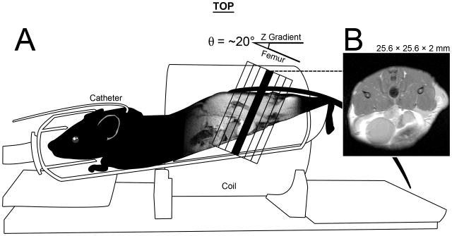
Figure 1.
An illustration of mouse positioning for MRI data acquisition. A) Mice were placed, prone and feet first, into a 38 mm inner-diameter, 33 mm long, 8 element, Doty-Litz coil. A custom mouse holder was built that elevated the hind limbs to minimize the angle θ between the laboratory z-axis and the angle of the femur. θ ranged from 12-25° and also served as the oblique angle for our axial images, which were perpendicular to the femur in the sagittal plane. The thigh muscle compartment rests above the lower abdomen, note the bladder in the ventral/anterior region of both sagittal and axial cross-sections. B) A representative image. All images were 25.6 × 25.6 × 2 mm. Multi-slice data (anatomical and DTI), had 7 slices that were centered on the femur (black outlined slices). Single slice data was acquired using the position of the center slice (black). Also shown is the placement of the jugular catheter used for DCE MRI.

