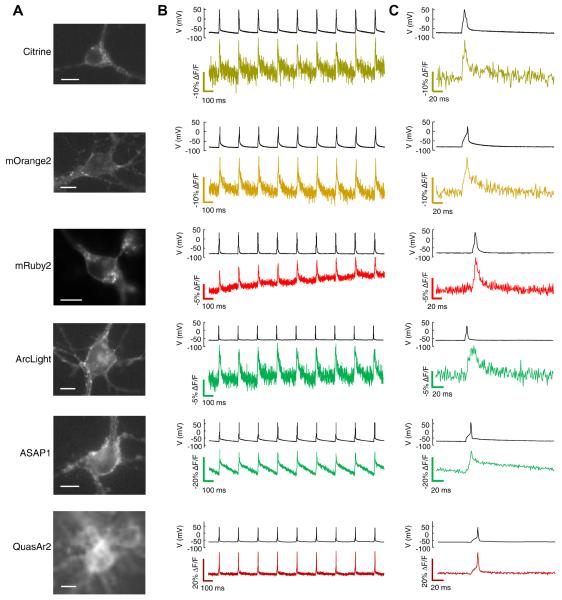
Figure 5. Single-trial recording of neuronal APs with GEVIs.
Each GEVI construct was expressed in primary rat hippocampal neurons under a CamKIIα promoter. Action potentials were induced by current injections through a patch pipette. A) Images of neurons expressing the indicated GEVI. Scale bars 10 μm. B) Simultaneous patch clamp and fluorescence recordings of AP waveforms. All recordings were acquired with laser illumination (3 W cm−2 for eFRET, ASAP1, and ArcLight GEVIs, 200 W cm−2 for QuasAr2) at an exposure time of 1 ms. Fluorescence traces are presented without temporal filtering nor correction for photobleaching. All but the QuasAr2 recording have been inverted (hence photobleaching appears as an upward trend in the mRuby2 trace). Recordings are representative traces from 5 – 11 cells recorded for each construct. C) Close-up showing single-trial, unfiltered, electrically and optically reported AP waveforms.

