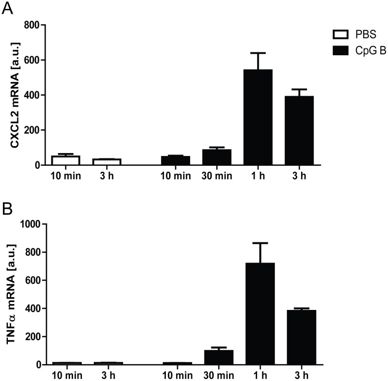
Figure 1. In vivo pharmacological assessments of TLR9-activation/inhibition.
Temporal profiles: Male C57BL/6 mice were injected i.p. with 100 µl 50 µg CpG B (black bars) or vehicle (white bars). Hearts were explanted at different time points (10 min, 30 min, 1 h and 3 h) and TLR9-mediated response was analyzed by mRNA expression levels of CXCL2 (panel A) and TNFα (panel B). Each data point represents mean ± SEM of 5 mice.

