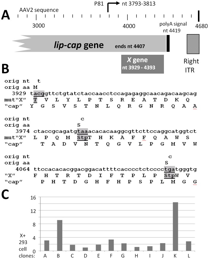
Figure 4. Environs of the X gene and reagents for X study.
A shows the region of X at the 3′ end of AAV2. Included are the 3′ end of lip-cap [1], [11], the p81 promoter [10], the poly A sequence and the 3′, right, inverted terminal repeat (ITR). B shows three nt substitution mutations in X which eliminate the products from all three 5′/amino end X start methionines, but which have no effect on the cap ORF/coding sequence. C shows the analysis of twelve 293 cell clones generated by transfection of pCI/X/Neo, and then G418 selected. The left scale shows the copy number of X found by Q-RT-PCR with clone D as the “1X” reference clone. 293-X-B and 293-X-K, having the highest copy numbers of X were chosen for further study.

