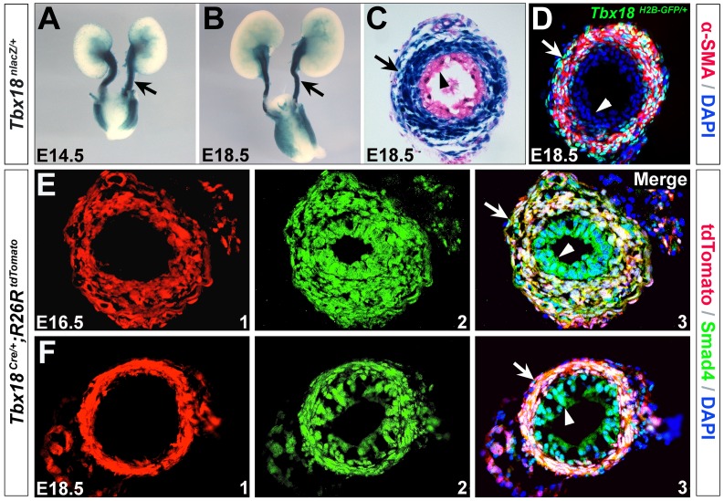
Figure 1. Expression and lineage analysis of Tbx18 in the developing ureters.
(A-C) Tbx18 expression in the developing ureters. A and B are whole-mount X-gal staining of Tbx18nlacZ/+ urinary system at E14.5 and E18.5, respectively. C is a transverse section of the ureter tube at E18.5. (D) Immunostaining of α-SMA (red) on Tbx18H2BGFP/+ ureter (transverse section) at E18.5. (E,F) Immunofluorescence analysis of Smad4 with Tbx18 lineage on Tbx18Cre/+;Rosa26tdTomato ureter at E16.5 and E18.5. Arrowheads indicate urothelium and arrows indicate ureter tube (A and B) or ureteral SMCs (C-F).

