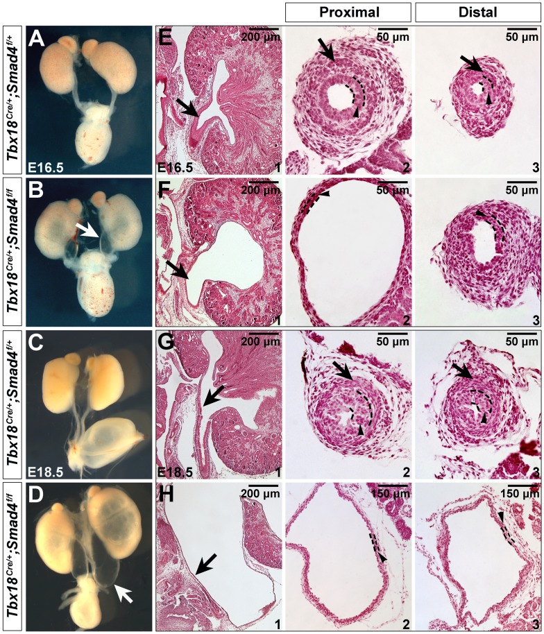
Figure 3. Disruption of Smad4 leads to hydroureter and hydronephrosis.
(A-D) Morphology of urinary system in the control and mutant mice at E16.5 (A and B) and E18.5 (C and D). (E-H) Hematoxylin and Eosin staining of kidney sagittal sections (E1/F1/G1/H1), and transverse sections of ureter tube in the proximal (E2/F2/G2/H2) and distal positions (E3/F3/G3/H3) at E16.5 and E18.5. Arrows in B/D and E1/F1/G1/H1 indicate ureter tube, and in E2/F2/G2/H2 and E3/F3/G3/H3 indicate ureteral SMCs. Arrowheads indicate urothelium.

