
Figure 2. Effect of phosphorylation on the DNA-Tau protein interaction.
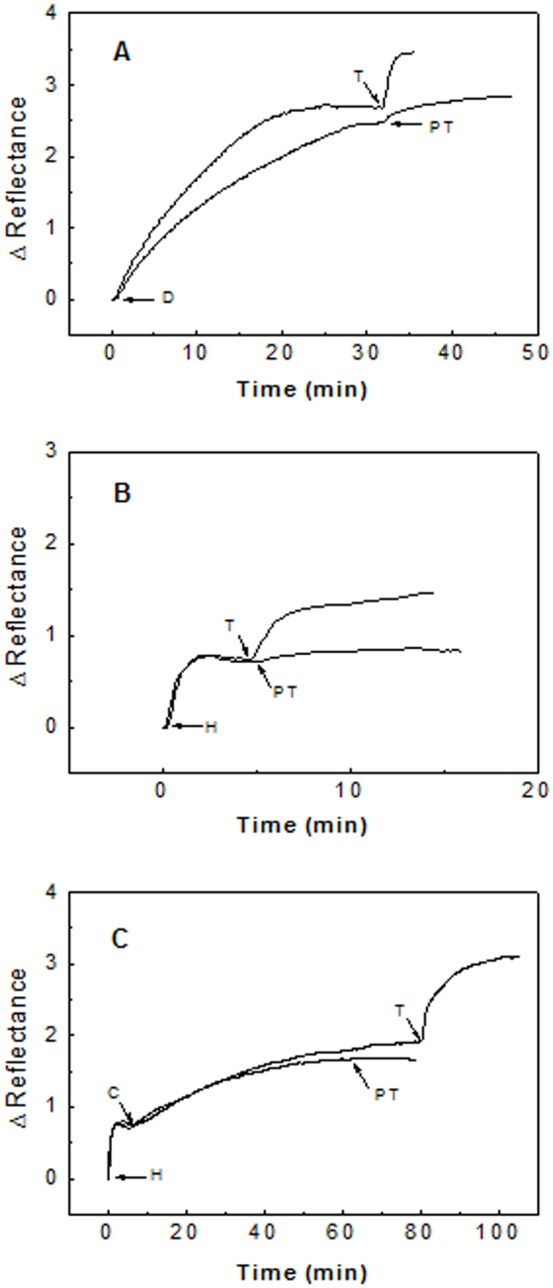
Part A: Tau protein (12 µg/mL) (T) and phosphorylated Tau (12 µg/mL) (PT) binding to a DNA activated sensor surface (D). Part B: Tau protein (10 µg/mL) (T) and phosphorylated Tau (10 µg/mL) (PT) binding to the heparin activated sensor surface (H). Part C: Tau protein and phosphorylated Tau binding to a DNA-histone mixture immobilized on the sensor surface. DNA (8 µg/mL) was incubated with histone (8 µg/mL) for one hour. At the time indicated by C, the incubated mixture was flowed on a heparin activated surface. Tau protein (T) and phosphorylated Tau (PT), both at 10 µg/mL, were then flowed through the sensor cell. All phosphorylated Tau forms were flowed during at least 10 minutes, which is enough for the full binding of Tau protein in Figures 2A and 2B, and for more than 60% in Figure 2C.