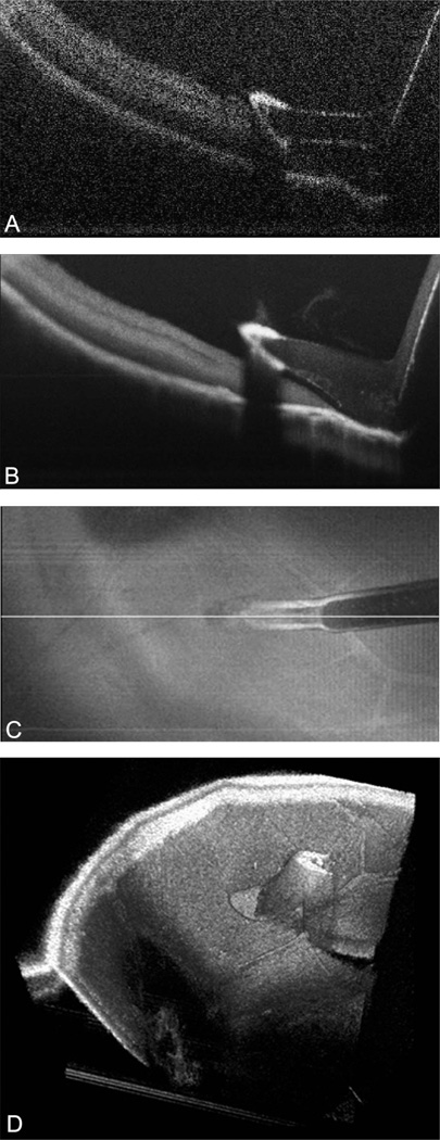
Fig. 1.
Microscope-mounted OCT image of diamond-dusted membrane scraper with cadaveric porcine retina. Image shows typical cadaveric porcine retina characteristics with high reflectance and minimal layering within retinal architecture. A. Single B-scan of diamond-dusted membrane scraper showing increased reflectance at area of diamond coating with increased shadowing. B. Summed B-scan series of diamond-dusted membrane scraper with improved tissue and instrument visualization and decreased shadowing under the silicone sleeve. Significant shadowing persists under the diamond coating. C. Summed voxel projection of diamond-dusted membrane scraper on retinal surface. D. Three-dimensional reconstruction of diamond-dusted membrane scraper on retinal surface.

