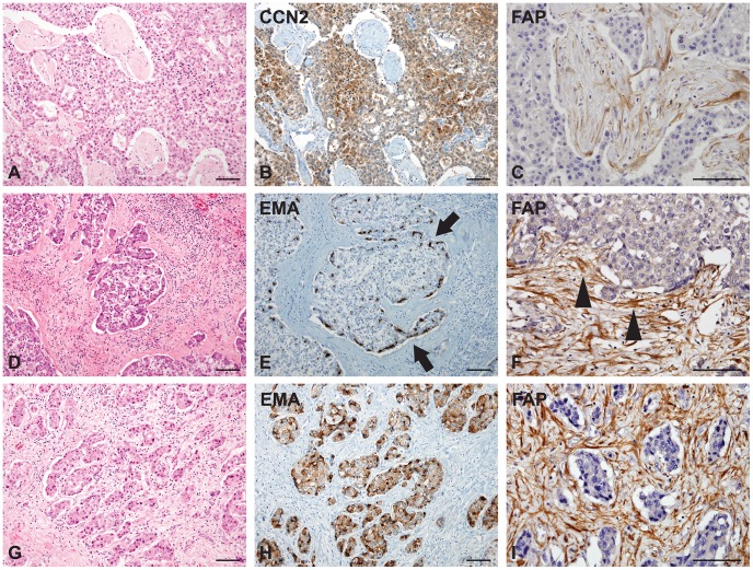
Figure 3. Connective tissue growth factor (CCN2), epithelial membrane antigen (EMA), and fibroblast activation protein (FAP) expression in scirrhous hepatocellular carcinomas (HCCs with abundant fibrous stroma) of cohort 2.
A–C) CCN2 (B) is diffusely expressed in the nests of tumor epithelial cells, and the tumor stromal cells between the nests of tumor epithelial cells exhibit strong FAP expression (C). D–F) EMA is mainly expressed in the periphery (E, arrows) of large tumor nests in contact with FAP-expressing cancer-associated fibroblasts (CAFs) of tumor fibrous stroma (F, arrowheads). G–I) HCCs with small nests or a trabecular pattern show diffuse expression for EMA in the tumor epithelial cells (H), which are closely admixed with FAP-expressing CAFs of tumor stroma. (Scale bars represent 100 µm.)

